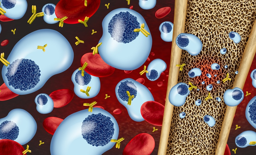
Etentamig in Relapsed/Refractory Multiple Myeloma: Pooled Phase 1 Data of an Investigational BCMA Bispecific Antibody
National Institutes of Health (NIH) | (.gov) (USA) · November 16, 2026, 11:37 PM
 mielomamultiplo.com · Multiple Myeloma
information · support · hope
It is always better to be an informed patient.
News, testimonies, a space to share ideas and links that help patients and carers understand the disease better and know where to find help.
Recent developments regarding the treatment and research of multiple myeloma indicate promising advances. [USA] A new bispecific antibody targeting BCMA is being tested in patients with relapsed multiple myeloma, while [UK] the inhibition of the NFIL3-MYC axis is emerging as a strategy to overcome therapeutic resistance. [Unidentified Country] New therapies, including CAR-T therapies and antibody combinations, are being approved, and FDA data analysis suggests the potential of Teclistamab with Daratumumab.
Made by MielomaAI On November 17, 2026, at 09:45, based on recent news. May contain errors. View news →
The plasmacytoma is the “physical” form of myeloma: instead of being scattered throughout the bone marrow, the diseased plasma cells clump together into a mass. It can be First stage of the diseaseAlone (solitary plasmacytoma, which is rare), or arising as a “child” of an existing myeloma.
It's a tumor made of plasma cellsBlood cells that are born in the bone marrow and produce antibodies (immunoglobulins) to defend the body from infections. In multiple myeloma, an altered plasmacyte multiplies uncontrollably and all copies make the same antibody. patients, all together in one place. It’s the “physical” form of myeloma: a mass that is seen on imaging tests and that, sometimes, can be felt by touch.
In multiple myeloma, the plasma cellsBlood cells that are born in the bone marrow and produce antibodies (immunoglobulins) to defend the body from infections. In multiple myeloma, an altered plasmacyte multiplies uncontrollably and all copies make the same antibody. patients are spreading bone marrowThe soft tissue inside the bones where blood cells are made: red blood cells, white blood cells, platelets, and plasma cells. It is examined with a bone marrow tap (bone marrow scan) or biopsy. It affects many bones. In the plasma cell tumorA tumor formed by plasma cells in one location. It can be in a bone (osseous) or in soft tissue, such as the nose or throat (extramedullary).They come together as one.
Spread out or in one placeIt originates within a bone, primarily in the vertebrae, pelvis, femur, or ribs. It can cause pain, fracture the bone, or compress the nerves of the spine.
The most common of the lonely onesIt originates outside the bones, in soft tissue: often in the nose, sinuses or throat, and more rarely in the stomach, intestines or lungs. It can cause a blocked nose, nosebleeds or hoarseness.
Out of the boneIn some patients, the plasma cell tumorA tumor formed by plasma cells in one location. It can be in a bone (osseous) or in soft tissue, such as the nose or throat (extramedullary). It's the first sign: it appears alone, before myeloma is present in the rest of the bone marrow. It's rare: in a large hospital, about 6 in every 100 patients with the disease plasma cellsBlood cells that are born in the bone marrow and produce antibodies (immunoglobulins) to defend the body from infections. In multiple myeloma, an altered plasmacyte multiplies uncontrollably and all copies make the same antibody. They had a solitary plasmacytoma.
First stage, in a few casesA biopsy of the mass is performed to see if they are plasma cellsBlood cells that are born in the bone marrow and produce antibodies (immunoglobulins) to defend the body from infections. In multiple myeloma, an altered plasmacyte multiplies uncontrollably and all copies make the same antibody. from a single clone. Then myeloma is looked for in the rest of the body: bone marrow biopsyBone marrow examination, almost always in the pelvis, with local anaesthesia: a small amount of bone marrow is aspirated (bone marrow aspiration/myleogram) and a small piece of bone is taken (bone biopsy)., analyses of protein MMonoclonal protein: the antibody (or antibody fragment) that is identically produced by myeloma cells. It is measured in the blood and urine and is used to see if the treatment is working. and a whole-body imaging scanPETPositron emission tomography (PET) scan that shows the areas of the body with very active cells. Helps to see lesions in the bones and disease outside the bone marrow.- CT or MRI.
Full-body examsIf there isn't plasma cellsBlood cells that are born in the bone marrow and produce antibodies (immunoglobulins) to defend the body from infections. In multiple myeloma, an altered plasmacyte multiplies uncontrollably and all copies make the same antibody. patients in the bone marrow, it's lonely. If there are a few, but less than 10%, it's called solitary with minimal bone marrow involvementWhen, in a solitary plasmacytoma, a few sick plasmocytes are found in the bone marrow, but less than 10%, the risk of progressing to myeloma is higher.And the risk of progressing to myeloma is quite a lot higher.
Change the riskO plasma cell tumorA tumor formed by plasma cells in one location. It can be in a bone (osseous) or in soft tissue, such as the nose or throat (extramedullary). Solitary myeloma is generally treated with radiotherapyTreatment with radiation directed to a specific area of the body. In myeloma, it is used in solitary plasmacytoma and to relieve bone pain. In the tumor area, every weekday for approximately 4 weeks. It often disappears. In the extramedularOutside the bone marrow: plasmacytoma tumors in organs or soft tissues (skin, liver, lymph nodes, nervous system). This indicates a more aggressive disease. You can also undergo surgery.
Around 4 weeksIn a patient who already has myeloma, new plasmacytomas can develop: attached to one bone, growing outside of it.bone marrowPlasma tumors that arise in a bone and grow outside of it into surrounding tissues carry a lower risk than free-floating tumors in organs.), or loose in other organsextramedullary diseaseMultiple myeloma with extramedullary deposits in organs or soft tissues, away from the bones (skin, liver, lymph nodes, nervous system). This indicates a more aggressive disease.They are like "sons" that the myeloma launches from the bone marrow.
In diagnosis or relapseLoose plasmacytomas, away from the bones—on the skin, liver, lymph nodes, or nervous system—indicate a more aggressive myeloma. They are treated with the general myeloma treatment, often more intense, and sometimes with radiotherapyTreatment with radiation directed to a specific area of the body. In myeloma, it is used in solitary plasmacytoma and to relieve bone pain..
High riskApproximately half of solitary bone plasmacytomas and around three in ten extramedullary plasmacytomas progress to myeloma within 10 years. Therefore, regular monitoring and imaging tests are carried out, even if the tumor has disappeared.
Regular follow-upA new pain in a bone, a lump that grows, weakness or tingling in the legs, or loss of control of urine or feces: contact your team or go to the emergency room immediately. There is hope, there is a way forward.
Don't wait for the appointment.Simplified explanation for patients and families. Only the hematology team can say what type of multiple myeloma it is and what the best treatment is. Source: Caers et al., Journal of Hematology & Oncology 2018 — recomendações europeias para o plasmocitoma solitário ↗ · This does not replace the explanation from your medical team.
It arises from the myeloma. plasma cellsBlood cells that are born in the bone marrow and produce antibodies (immunoglobulins) to defend the body from infections. In multiple myeloma, an altered plasmacyte multiplies uncontrollably and all copies make the same antibody., as células da bone marrowThe soft tissue inside the bones where blood cells are made: red blood cells, white blood cells, platelets, and plasma cells. It is examined with a bone marrow tap (bone marrow scan) or biopsy. What antibodies do. Not all multiple myeloma are the same: they change phase, the protein they produce and the risk.
There is plasma cellsBlood cells that are born in the bone marrow and produce antibodies (immunoglobulins) to defend the body from infections. In multiple myeloma, an altered plasmacyte multiplies uncontrollably and all copies make the same antibody. modified and a little abnormal protein in the blood (a) protein MMonoclonal protein: the antibody (or antibody fragment) that is identically produced by myeloma cells. It is measured in the blood and urine and is used to see if the treatment is working.), but without symptoms or damage. It's not cancer and it's not treated: it's monitored with tests.
Progresses slowly: about 1% per yearMore protein MMonoclonal protein: the antibody (or antibody fragment) that is identically produced by myeloma cells. It is measured in the blood and urine and is used to see if the treatment is working.Or 10% or more plasma cellsBlood cells that are born in the bone marrow and produce antibodies (immunoglobulins) to defend the body from infections. In multiple myeloma, an altered plasmacyte multiplies uncontrollably and all copies make the same antibody. in the bone marrow, but without damage to the organs. Most cases are monitored; in latentSmoldering myeloma: protein M of 3 g/dL or higher, or 10% to 60% plasma cells in the bone marrow, but without organ damage. Approximately 10% per year evolve to active myeloma in the first 5 years. High risk may already be treatable.
Close monitoringWhen myeloma has already caused damage – high calcium, kidneys affected, AnemiaLack of red blood cells or hemoglobin in the blood. Causes fatigue, paleness, and shortness of breath. or bone injuries (the signsCRABThe English acronym for the 4 typical damages of myeloma: C for high calcium in the blood, R for kidneys (kidney failure), A for anemia, and B for bone, i.e., lesions in the bones. Just one of these is enough to speak of active myeloma.) — or when tests show it’s going to cause it soon. Then treatment is needed.
It needs to be treated.Each antibody has 2 heavy chainsThe larger parts of an antibody. There are 5 types – G, A, D, E and M – and they are what give the immunoglobulin its name: IgG, IgA, IgD, IgE or IgM. e 2 fusion clampsThe smallest fragments of an antibody. There are two types, kappa and lambda. They are measured in the blood using the free light chain test; a significantly altered kappa/lambda ratio indicates a plasma cell clone.. Multiple myeloma is named after the heavy chain (G, A, D, E or M) and the light chainkappaOne of the two light chain types. Approximately 2 in every 3 multiple myeloma cases are kappa. or That's a good question.One of the two light chain types. Approximately 1 in every 3 multiple myeloma cases is lambda; it is also the most common type in AL amyloidosis.:) for example, “IgG kappa”.
Discovered with immunofixationIgG is the most common: more than half of patients. IgA follows, and more often forms tumors outside of the bones. The treatment is generally the same.
The most commonApproximately 1 in every 5 patients only produces fusion clampsThe smallest fragments of an antibody. There are two types, kappa and lambda. They are measured in the blood using the free light chain test; a significantly altered kappa/lambda ratio indicates a plasma cell clone. It (the protein) Bence JonesOld term for light chains found in urine. It comes from the English doctor who described them in the 19th century.). They are small and can block the kidneys, which need extra attention. Followed by with... free catenaryLoose pieces of antibody floating in the blood. The two types (kappa and lambda) are measured, as well as the ratio between them; a very high alteration indicates a clone of plasma cells. in the blood.
Around 20% of patientsIgD, IgE and IgM are rare. In smoldering myeloma (1-2% of patients), there is almost no protein detected in the blood tests: follow-up with bone marrow examination and imaging tests, such as PETPositron emission tomography (PET) scan that shows the areas of the body with very active cells. Helps to see lesions in the bones and disease outside the bone marrow..
RareA single tumor plasma cellsBlood cells that are born in the bone marrow and produce antibodies (immunoglobulins) to defend the body from infections. In multiple myeloma, an altered plasmacyte multiplies uncontrollably and all copies make the same antibody.It can be in a bone, or outside of one, without myeloma in the rest of the bone marrow. It is often treated with radiotherapyTreatment with radiation directed to a specific area of the body. In myeloma, it is used in solitary plasmacytoma and to relieve bone pain. and then you are monitored, as it can progress.
Radiotherapy and monitoringSome changes in the chromosomes of myeloma cells make the disease more aggressive: loss of 17pA fragment of chromosome 17 where the TP53 gene is located, which controls cell growth. If it is lost (17p deletion), the myeloma tends to be more aggressive. or mutation TP53Gene that acts as a brake on cells. When it is altered (mutated or lost), the myeloma responds worse to some treatments., e t(4;14)Translocation: exchange of pieces between chromosomes 4 and 14. It doesn't count as high risk on its own; it does count when it's associated with gains in 1q or losses in 1p.t(14;16) or t(14;20) when joining to gain 1qLong arm of chromosome 1. Having extra copies (a gain of 1q) is common and makes the disease more difficult to control, especially when combined with other changes. or a loss of 1 point. Two or more: even greater risk.
High riskA Multiple myelomaRare and very aggressive condition: there are 5% or more plasma cells circulating in the blood. It can occur at the onset (primary) or as a relapse of myeloma (secondary). (5% or more) plasma cellsBlood cells that are born in the bone marrow and produce antibodies (immunoglobulins) to defend the body from infections. In multiple myeloma, an altered plasmacyte multiplies uncontrollably and all copies make the same antibody. (circulating in the blood) and the extramedullary diseaseMultiple myeloma with extramedullary deposits in organs or soft tissues, away from the bones (skin, liver, lymph nodes, nervous system). This indicates a more aggressive disease. Extramedullary disease is the most aggressive form.
Very high riskToday, a combination of 4 medications is often used: one anti-CD38Antibodies (daratumumab, isatuximab) that bind to the CD38 protein, which is very prevalent in myeloma cells, and mark them for destruction by the immune system. (daratumumab), a proteasome inhibitorMedications (bortezomib, carfilzomib, ixazomib) that block the protein “breaker” in the cell. The myeloma cells, which produce a lot of protein, accumulate waste and die. (bortezomib), a immunomodulatorTablets (lenalidomide, pomalidomide, talidomide) that inhibit myeloma and help the body’s defenses attack it. lenalidomide and a corticosteroidCorticosteroid medications, such as dexamethasone. At high doses, they kill myeloma cells and enhance the effect of other medications. (dexamethasone)
Introduction: several monthsWho can, does. allogeneic transplantStem cells are collected from the patient's own body, followed by intensive chemotherapy (melfalan) and then the cells are returned to the bone marrow to recover.And it involves using your own stem cells. Following this... MaintenanceMilder and longer treatment after induction and transplant, to keep the myeloma under control for a longer period. Often it is lenalidomide in tablet form.Generally, with lenalidomide, to keep the disease under control.
Transplant and maintenanceThere are other weapons: new combinations, antibodies. Specific biomarkersAntibodies with two “arms”: one grabs the T lymphocyte and the other the myeloma cell, joining them so that the lymphocyte destroys. Examples: teclistamab, elranatamab, talquetamab. e CAR-TTreatment involving the patient’s own T lymphocytes, modified in the laboratory to recognize the myeloma and then reintroduced through infusion.In high-risk myeloma, doctors often treat more intensively and may propose clinical trialsStudies comparing a new treatment with the best existing treatment may provide access to medications before approval. Participation is voluntary..
RelapseIn all types: medications to strengthen bones, vaccines and prevention of infections, and attention to the kidneys are part of the treatment.
AlwaysUnder the film is the complete list: each type, what it is, how it progresses or is treated, and the risk, including related diseases, such as Amyloidosis ALDisease in which the light chains misfold and deposit themselves in the organs (heart, kidneys, nerves), damaging them. It can exist on its own or with multiple myeloma. e a POEMSRare syndrome linked to a plasma cell clone (almost always lambda): Polyneuropathy, Organomegaly, Endocrinopathy, M Protein and Skin Changes..
Movie listings belowEach myeloma is different. Ask your team: what is my type, what stage am I in, and what is my risk? There is hope, there is a path.
Simplified explanation for patients and families. The type, stage and risk for each person can only be determined by the hematology team. Source: International Myeloma Foundation — Types of Myeloma ↗ · This does not replace the explanation from your medical team.
Click on a type to open. Hover over (or tap) the underlined words to see what they mean.
The risk doesn't just depend on the type. Doctors use the R-ISS or R2-ISS stage… beta-2 microglobulinProtein measured in the blood. High values indicate more disease and are included in the staging (ISS) calculation.The albumin, the LDHMeasured enzyme (lactate dehydrogenase). High values may indicate more active disease. and genetics) and are looking for high-risk changes: loss of 17pA fragment of chromosome 17 where the TP53 gene is located, which controls cell growth. If it is lost (17p deletion), the myeloma tends to be more aggressive. or mutation TP53Gene that acts as a brake on cells. When it is altered (mutated or lost), the myeloma responds worse to some treatments.It’s a condition where parts of your chromosomes swap places. t(4;14)Translocation: exchange of pieces between chromosomes 4 and 14. It doesn't count as high risk on its own; it does count when it's associated with gains in 1q or losses in 1p.t(14;16) or t(14;20) alongside gains 1qLong arm of chromosome 1. Having extra copies (a gain of 1q) is common and makes the disease more difficult to control, especially when combined with other changes. or a loss of 1 point, and very high beta-2 microglobulin with normal kidneys. Two or more changes = an even greater risk.
| Type | Where | How frequent | Treatment | Risk of myeloma |
|---|---|---|---|---|
| Solitary bone plasmacytoma | Inside a bone: vertebrae, pelvis, femur, ribs. | Approximately 4 in 100 patients with plasmacytosis disease. | RadiotherapyTreatment with radiation directed to a specific area of the body. In myeloma, it is used in solitary plasmacytoma and to relieve bone pain. (40 to 50 Gy, approximately 4 weeks) | Approximately half progress to myeloma within 10 years. |
| Solitary extramedullary plasmacytoma | Outside the bone: nose, sinuses, throat; less commonly stomach, intestines, lungs. | About 2 in every 100. | RadiotherapyTreatment with radiation directed to a specific area of the body. In myeloma, it is used in solitary plasmacytoma and to relieve bone pain.Sometimes surgery. | About 3 in 10 progress to the advanced stages within 10 years. |
| Solitary with minimal involvement of the bone marrow | It plasma cell tumorA tumor formed by plasma cells in one location. It can be in a bone (osseous) or in soft tissue, such as the nose or throat (extramedullary). and less than 10% plasma cellsBlood cells that are born in the bone marrow and produce antibodies (immunoglobulins) to defend the body from infections. In multiple myeloma, an altered plasmacyte multiplies uncontrollably and all copies make the same antibody. Patients in the bone marrow. | Monitor closely when using sensitive techniques (flow cytometry). | RadiotherapyTreatment with radiation directed to a specific area of the body. In myeloma, it is used in solitary plasmacytoma and to relieve bone pain. and closer monitoring; sometimes a clinical trial. | Much larger: in a study, 7 out of 10 progressed (compared to less than 1 in 10 without affected marrow). |
| Vertebral plasmacytomas (in multiple myeloma) | They are born in a bone and grow into the surrounding tissues. | Relatively common in the diagnosis of multiple myeloma. | General myeloma treatment radiotherapyTreatment with radiation directed to a specific area of the body. In myeloma, it is used in solitary plasmacytoma and to relieve bone pain. If you're feeling anxious or in pain. | Falls under myeloma; intermediate risk. |
| Extramedullary disease (in myeloma) | Afflictions in organs distant from the bones: skin, liver, lymph nodes, nervous system. | Rare in the diagnosis; more common in relapses. | Intensive treatment CAR-TTreatment involving the patient’s own T lymphocytes, modified in the laboratory to recognize the myeloma and then reintroduced through infusion. e Specific biomarkersAntibodies with two “arms”: one grabs the T lymphocyte and the other the myeloma cell, joining them so that the lymphocyte destroys. Examples: teclistamab, elranatamab, talquetamab. in relapse clinical trialsStudies comparing a new treatment with the best existing treatment may provide access to medications before approval. Participation is voluntary.. | High risk: more aggressive disease. |
Numbers of the European recommendations for solitary plasmacytomaCaers et al., 2018 ↗Gray (Gy) is the unit of radiation therapy dose. Each case is assessed by the hematology and radiation therapy team.

Comprehensive and up-to-date news coverage, gathered from sources around the world by Google News.

Ben Derman: Top 10 Myeloma Abstracts to Be Presented at ASH 2026 / ASCO, ASH 2026, Ben Derman, cancer, CAR-T, DARA, Daratumumab, Lenalidomide, Mount Sinai

MRD is becoming a very valuable tool, but it's not an exclusive tool, says expert
All news → · Summaries created by MielomaAI; expand the article to read the original.

The symbol brings together two stories: that of the macedotuga.com, from Vila do Conde, and that of multiple myeloma.
It is always better to be an informed patient. There is hope. There is a way forward.